Booking inPune - 411005

Cart


Browse by Categories

Full Body Checkup
Overall health diagnosis
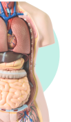
Body Organs
Assess major organ function

Lifestyle Habits
Measure habits, empower health

X-Ray, MRI & Scans
Radiology based lab tests
What is a USG test?
An ultrasound is a medical imaging test. It uses sound waves to create detailed pictures of organs, tissues, and other structures inside the body. It is a non-invasive procedure that helps doctors visualize and diagnose various conditions without surgery. The images produced are called sonograms.
Other names of USG scan:
-Ultrasound -Ultrasonography -Sonography -Diagnostic ultrasound imaging -Ultrasound imaging -pregnancy sonography
Significance of the USG test:
Use to monitor the health and development of an unborn baby during pregnancy. - Useful in guiding certain biopsy procedures. - Helps diagnose gallbladder disease - It can help assess the thyroid gland for abnormalities, including nodules or enlargement. - Helps visualize breast tissues and can assist in identifying breast abnormalities, like cysts or solid masses.
How does a USG scan work?
A healthcare professional called a sonographer uses a wand-like device called a transducer, which emits high-pitched sound waves. The transducer is moved gently over the skin. A special gel is applied to help transmit the sound waves and ensure good contact between the skin and the transducer. The applied gel also helps reduce air pockets that can interfere with sound wave transmission. The transducer emits high-frequency sound waves into the body, which then bounce back from different tissues and structures. The emitted sound waves are captured by the wand (transducer) and converted into electrical signals. A computer then processes the collected signals to create real-time images that are displayed on a monitor.
Types of USG:
Transesophageal echocardiogram: Uses a transducer inserted into the esophagus to obtain heart images (usually done under sedation). - Transrectal ultrasound: Creates images of the prostate by placing a transducer into the rectum. - Transvaginal ultrasound: In this transducer is gently inserted into the vagina to examine the uterus and ovaries.
Preparation for a USG:
Preparation depends on the body part being examined and will be communicated by your healthcare provider. - Some ultrasounds may require no preparation, while others may involve fasting or drinking water to have a full bladder.
Benefits and risks of USG scan:
Benefits of USG: - Non-invasive: USG does not involve any incisions or radiation exposure, making it a safe imaging option. - Real-time imaging: USG provides real-time images, which help doctors to observe and evaluate structures and functions of the internal organs. - Versatile: USG can be used to examine various parts of the body, making it a valuable diagnostic tool in different medical fields. - Safe for most individuals: USG uses sound waves and not radiation to examine the organs. Therefore, it is generally safe for people of all ages, including infants, and pregnant women. Risks and limitations: No known significant risks associated with ultrasound imaging. However, like all other medical procedures, USG has a few limitations. The test may not provide detailed images of certain structures, like those located deep within the body or obscured by bone. In such cases, other imaging modalities like MRI or CT scans may be more appropriate.
What to expect during USG test?
A sonographer will apply a special gel on the skin over the area that needs to be examined. - The transducer will be gently moved over the skin to capture the images. - The exam typically takes around 30 minutes to an hour, depending on the test and the body part being examined.
What to expect after USG?
After the exam, the sonographer will wipe off the gel from your skin. - There are usually no special restrictions or after-effects, and you can resume your normal activities.

How much does a scan cost in Chennai?
If you’re looking for affordable scan options in Chennai, Bajaj Finserv Health offers a wide range of diagnostic centers with transparent pricing. The cost of a scan can vary depending on the facility and the type of scan. We make it easy to compare prices from different labs so you can find the best option without compromising on quality. Book your scan online and get the best rates in Chennai today!
Where can I find a scan center near me?
Need to find a scan center near you? With Bajaj Finserv Health, you can easily locate trusted diagnostic centers in Chennai for your scan. Our platform connects you with certified labs in your area, ensuring that you receive high-quality service and accurate test results, right when you need them. Simply enter your location, and we’ll show you the best options for scan near you.
What is the price of a scan?
Wondering about the price of a scan? The cost of diagnostic tests like scan can differ depending on the facility, technology, and location. At Bajaj Finserv Health, we partner with top labs across Chennai to offer competitive pricing, giving you the flexibility to choose the lab that suits your budget without sacrificing quality. Explore options and book your test online for peace of mind.
Factors Affecting Scan Price in Chennai
- Lab certification and accreditation status
- Center location and accessibility
- Test methodology and equipment used
- Turnaround time for results
How much is a scan in Chennai?
The price of a scan in Chennai typically ranges based on the lab’s services and equipment. Bajaj Finserv Health partners with multiple diagnostic centers across the city, making it easy for you to find a reliable scan at an affordable cost. Whether you’re looking for standard or advanced tests, we ensure transparent pricing and convenient online booking, so you know exactly what to expect.
How can I book a scan in Chennai?
If you need a scan in Chennai, Bajaj Finserv Health offers a hassle-free booking process. You can find certified labs near you offering scan and other diagnostic services with just a few clicks. Whether it’s for a routine check-up or a specific medical need, our platform helps you find the right lab at the right price in Chennai.
How do I find a scan near me?
Searching for scan centers near you? Bajaj Finserv Health provides an easy-to-use platform to find and book diagnostic tests in your area. Whether you’re in Chennai or another location, you can locate top-rated labs offering scan tests at competitive rates. Simply enter your location, choose a lab, and book your test online for a seamless experience.
Why Choose Our Partner Labs for Scan in Chennai?
Our network of trusted diagnostic centers in Chennai ensures reliable scans with consistent quality. Enjoy added benefits such as home sample collection, quick access to digital reports, and affordable pricing.
Key Features
- Accredited NABL/ISO certified laboratories
- Access to digital reports for convenience
- Easy online booking and flexible payment options
How much does scan cost in Chennai?
The cost of scan in Chennai can vary based on the diagnostic center, the type of scan required, and additional factors like the use of contrast material. To get the best deal, visit our platform to check the latest prices and compare offerings from multiple accredited diagnostic centers
How can I find scan centers near me in Chennai?
Visit the Bajaj Finserv Health website and explore the Lab section to find accredited centers near you. You can compare prices and book appointments conveniently.
What factors affect scan prices in Chennai?
scan prices in Chennai vary based on scan complexity, equipment quality, use of contrast material, and additional services like consultations and report delivery.
What should I look for in a center in Chennai?
Choose a center with NABL accreditation, modern equipment, experienced radiologists, and digital report delivery for reliable results.
How can I book scan online?
You can book through our website or app, selecting your preferred center and time slot
What is the cost of scan in Chennai?
Prices start from ₹{{min_price}} and vary by center location and facilities.