Health Tests | 13 min read
Bone Density Test: Purpose, Procedure, Results, Risk Factor
Medically reviewed by
Table of Content
Key Takeaways
- Bone density test helps determine the minerals present in a bone segment
- DEXA scan is one the most accurate lab tests to measure bone density
- Bone density test can help your doctor diagnose osteopenia or osteoporosis
Your bones protect your organs and internal muscles which is why it is important to have good bone density. Apart from this, bones also help store calcium and give structure. Bone density refers to the number of minerals present in a certain volume of bone. A good bone density test indicates that your bones are strong, healthy, and less likely to break.
Your bones change continuously, meaning that old bones break and new bones form. This change is quicker when you are young, and you reach your peak bone mass around the age of 30 [1]. After this age, your bones keep changing but you may gain less bone mass than you lose. Apart from age, gender also plays a major role in the changes in bone density and bone problems. Women over the age of 50 are 4 times more likely to develop osteoporosis and 2 times more likely to develop osteopenia [2].
One of the ways you can check the health of your bones is through a bone density test. Also known as the bone mineral density test, this test helps determine how many minerals are present in a segment of bone. Read on to know more about bone density test, their purpose, and what the test results indicate.
What Is A Bone Density Test And Why Is It Done?
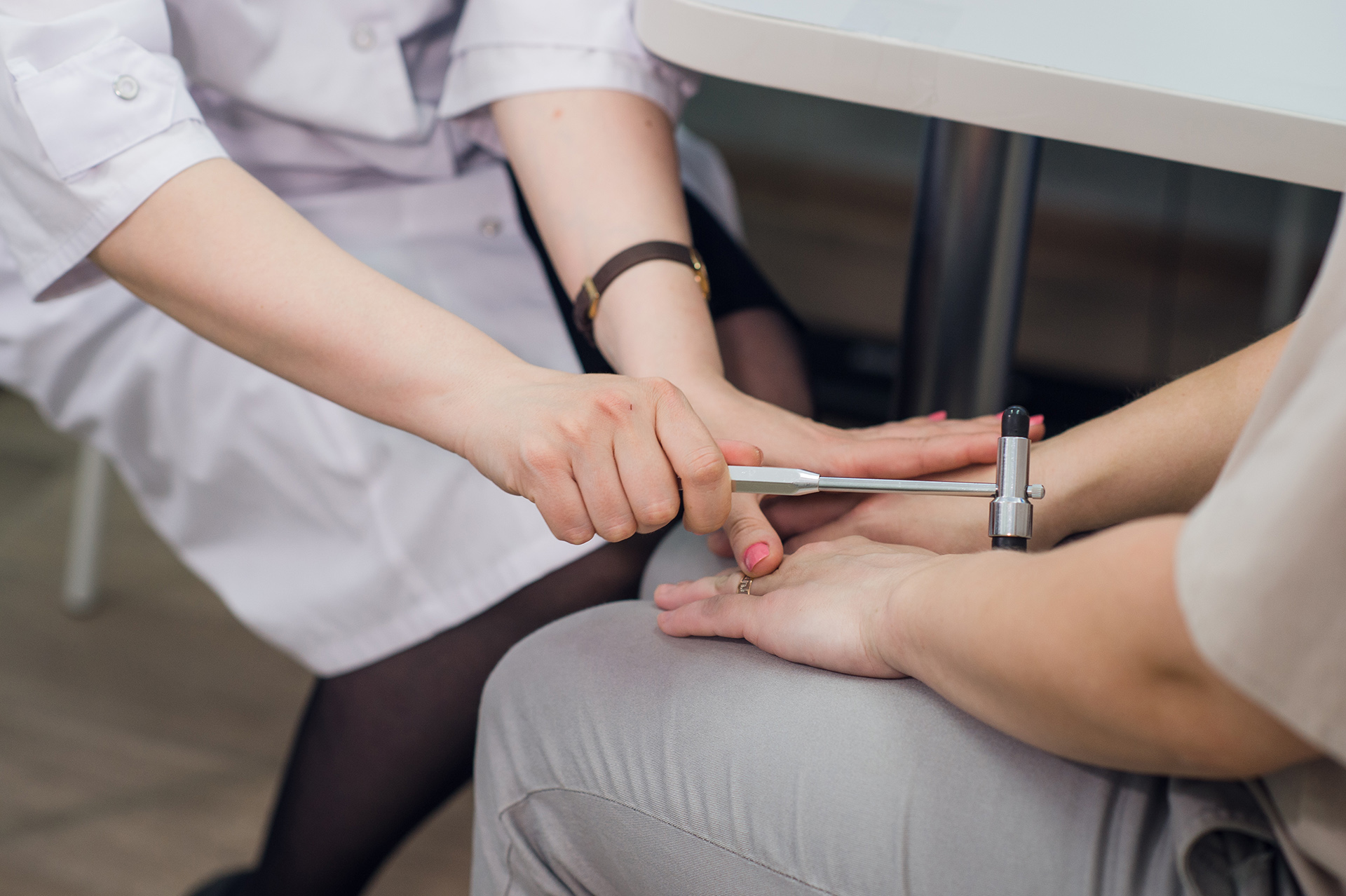
A bone density test is a lab test that helps determine whether you have osteoporosis. Bone density test is quick, painless, and performed with an X-ray. It measures the number of minerals present in a bone segment. It can also help recognize a decline in bone density and determine your risk of broken bones. Apart from this, a test can help keep track of your osteoporosis treatment.
Additional Read: Bone Marrow Biopsy
What Is A DEXA Scan?
A DEXA scan is a particular kind of imaging examination. A very low dose of x-rays is used to gauge how solid your bones are. Dual-energy X-ray absorptiometry is known as DEXA.
Medical professionals believe that DEXA scans are the most effective, convenient, and affordable diagnostic for identifying osteoporosis. In addition, the exam is painless and fast.
Risk Factors That Affect Bone Health
Family background
Bone health issues may run in the family. So first, check to see whether anyone in your family, especially your parents or siblings, has ever had osteoporosis diagnosed. This involves parents or siblings who have fractured a bone (from a small fall) or have grown shorter quickly, as these signs may point to osteoporosis risk.
Vitamin D and calcium
- Low calcium intake: Adults need 1,000 mg of calcium daily, ideally from their food, while men and women over the age of 70 need 1,300 mg daily
- Low vitamin D levels: Calcium absorption requires vitamin D. Low vitamin D levels can be caused by lack of sun exposure. Investigating those at risk of vitamin D insufficiency is essential
Medical background
Conditions and drugs that may influence bone health include:
- Anyone 50 years or older who breaks a bone after a small bump or fall should have it looked at
- Early menopause in women or low testosterone in males are symptoms of low hormone levels
- Inflammatory bowel disease, coeliac disease, and other malabsorption diseases
- Diabetes, prostate cancer, certain breast cancer treatments, or nervous anorexia
- Commonly prescribed corticosteroids for rheumatoid arthritis, asthma, and other inflammatory diseases
- Overactive or parathyroid conditions
- Rheumatoid arthritis
- Continual kidney or liver disease
- Some antidepressants, treatments for epilepsy, or HIV
Physique and weight:
- A lean physique might enhance your danger
- Studies show that obesity-related hormone alterations might influence bones
Lifestyle elements
- Insufficient physical activity
- Smoking
- Excessive alcohol consumption
What to Expect During Bone Density Test
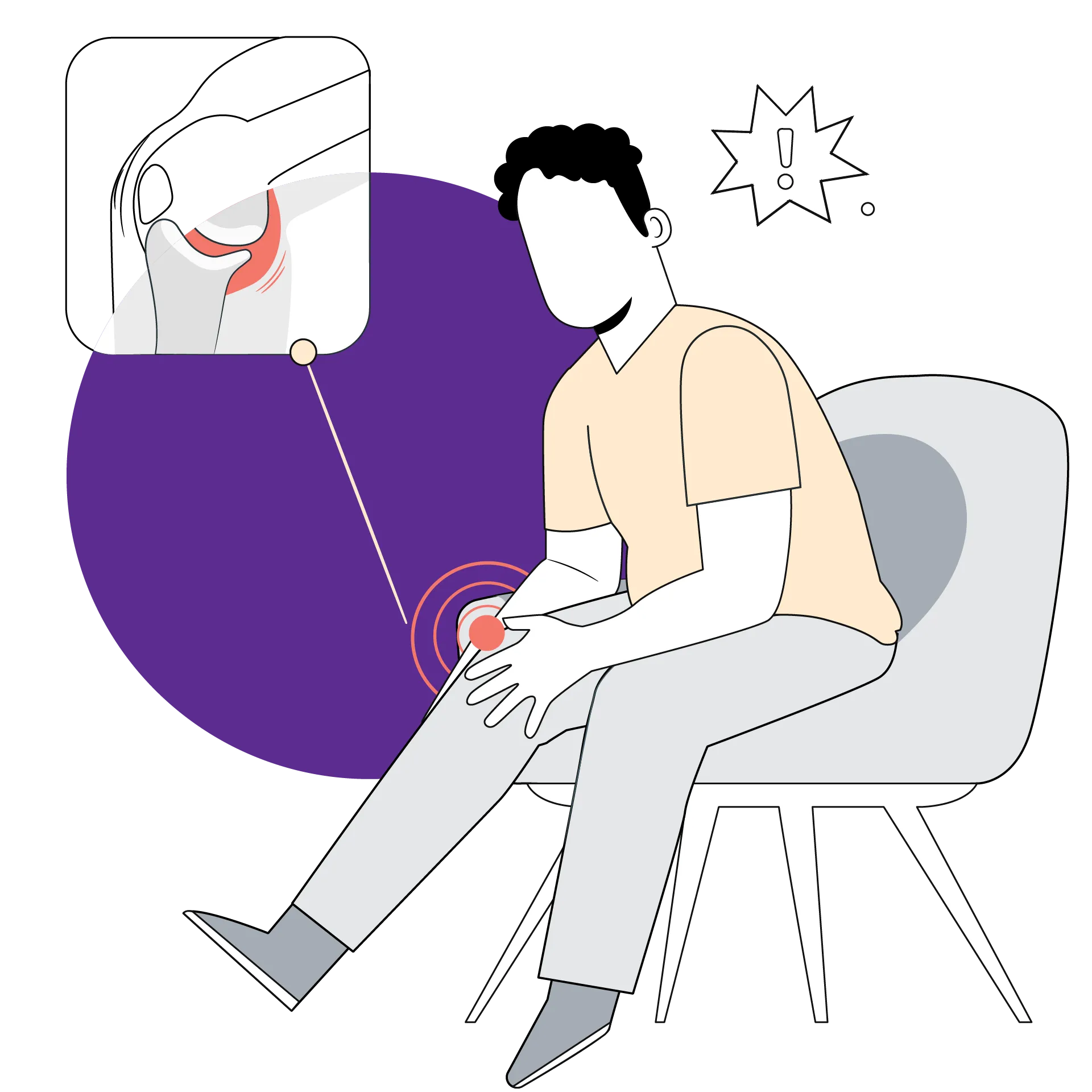
Typically, the examination analyses the joints in your vertebrae, hip, and forearm. When you have osteoporosis, the joints mentioned above are most prone to breaking.
Both sorts of bone density examinations take just under 15 minutes. They are:
Central DXA:
This examination examines your vertebrae and hip bones. It is more accurate. It also costs extra. Dual Energy X-ray Absorptiometry is referred to as central DXA.
During the exam, you lie fully dressed on a cushioned platform. A machine arm travels over you, transmitting low-dose X-rays into your body. It generates a picture of your skeleton depending on the number of X-rays that change after going through your bones. This exam lasts roughly 10 minutes. Then, an expert is given the photograph and reads the outcomes. This may need a few days, depending on your physician's office.
Test on the periphery:
This gauges the strength of your wrist, finger, and heel. Due to the absence of a spine or hip examination, this test is less comprehensive. Typically, this is cheaper.
Since the gadget is portable, it may be taken to pharmacies and health fairs. More people who would not be able to receive the central DXA test can opt for this one.
Peripheral tests are another method of screening patients, allowing those with a higher risk of osteoporosis to undergo further testing. They are also utilized for bigger individuals who cannot receive central DXA due to weight restrictions.
How to Prepare for Bone Density Test
- Avoid taking calcium supplements twenty-four hours before the assessment
- Wait seven days before getting a central DXA if you have a barium or contrast dye injection for a CT scan or MRI. Your bone density test might be affected by the contrast dye.
- Wearing clothing with metal belts, buttons, or zippers is not recommended.
Taking the exam carries very little risk. Your radiation exposure is far lower than that of a chest X-ray or a trip.
Who Should Get Bone Density Test
Osteoporosis may affect everyone. Men can also get it, but older women seem to have it more frequently. As you become older, your odds grow.
You should talk to your doctor about the test if necessary. They could advise if you fit any of the following criteria:
- You are at least 65 years old (female)
- 50 or older and a postmenopausal lady
- When a woman enters menopause, her risk of fracturing bones increases
- You have other factors that increase your risk of osteoporosis since you are a woman under 65 who has previously experienced menopause
- You're a male over 50 with additional risk factors
- You shatter a bone after the age of 50
- Your adult height has diminished by more than 1.5 inches
- Your stance has become more stooped
- You get back discomfort for no apparent reason
- Although you are neither pregnant nor menopausal, your periods have ceased or are erratic
- You underwent organ transplantation
- Levels of your hormones have fallen
Bone loss may result from using some prescription medications. Glucocorticoids, a class of drugs designed to reduce inflammation, would be one of these. If you have ever taken cortisone (Cortone Acetate), dexamethasone (Baycadron, Maxidex, Ozurdex), or prednisone, let your doctor know (Deltasone).
How Is A Bone Density Test Performed?
A flat, wide X-ray table is where you lie on your back for a DEXA scan. To prevent blurry images during the scan, you must remain steady.
Typically, a radiographer specializing in capturing X-ray pictures will do the scan.
A sizable scanner arm will be moved over your body throughout the scan to evaluate bone density in the middle of the skeleton.
A small beam of low-dose X-rays will be sent across the area of your body being scanned while the scanning arm is progressively moved over your body.
Your hip and lower spine will be examined for weak bones (osteoporosis). However, because bone density varies across the skeleton, more than one area of your body could be examined.
If scans of the hip or spine are not possible or necessary due to certain medical conditions, such as hyperparathyroidism, the forearm may be examined instead.
Your body will absorb X-rays administered through fat and bone tissue.
An X-ray detector inside the scanning arm counts the number of X-rays travelling through your body. This data will be needed to create a picture of the scanned region.
Typically, the scan takes ten to twenty minutes. Once everything has been completed, you will be allowed to return home.
There are various ways to measure your bone density. One most accurate and common way is the dual-energy X-ray absorptiometry (DEXA scan). It is usually performed in a radiologist’s lab. Your doctor may ask you to stop consuming calcium supplements 24-48 hours prior to the test.
There are various ways to measure your bone density. One most accurate and common way is the dual-energy X-ray absorptiometry (DEXA scan). It is usually performed in a radiologist’s lab. Your doctor may ask you to stop consuming calcium supplements 24-48 hours prior to the test.
Before the bone density test or scan, the doctor will ask you to lie on a padded table with your legs straight. A scanning machine will pass over your hips and lower spine. Another scanner, known as photon generator will pass from beneath you. The images from these two scanners are then sent to computer. You will need to stay very still during the scanning and you may also be asked to hold your breath for accurate results. The test may last for 10-30 minutes.
To measure the bone density in your hand, foot, or forearm, doctors use a portable scanner known as p-DEXA (peripheral DEXA).
Can DXA be Used for More Than Measuring Bone Density?
Beyond just assessing bone density, DXA may be used to assess your overall bone health. Here are a few further DXA applications. Some DXA facilities offer these tests, although not all of them.
Assessment of vertebral fractures (VFA):
This sideways picture of the spine can be used to identify fractures or crushed vertebrae. The majority of persons who suffer from these fractures are unaware of them. Your diagnosis, the assessment of your fracture risk, and your treatment options may alter if a previously undiagnosed spine fracture is found.
Trabecular Bone:
The trabecular bone score (TBS) is a measurement of the microscopic internal organization of the bones in your spine. The better it is, the higher the number. It is produced using specialized software that is integrated into the DXA system. The TBS number can be added to FRAX to provide a more accurate evaluation of fracture risk.
Full-length femur Imaging:
FFI or full-length femur imaging FFI is a method for using DXA to obtain a complete picture of your femur (thigh bone), as opposed to simply the region surrounding the hip, which is visible with conventional DXA. This can help identify bone thickening that can cause a stress fracture or an unusual femur fracture.
Hip structural analysis (HSA):
Your hip's strength and the chance of breaking can be affected by the size, form, and arrangement of its bones. HSA with DXA offers a perspective on this and may occasionally aid in making treatment options.
Are There Other Tests For Bone Density And Bone Health?
DXA is only one of several tests that may be done to evaluate your bone health. While some of them are less often used than DXA, they may offer important information that goes beyond bone density or even assist in deciding who requires a DXA.
Computed tomography in quantity (QCT)
QCT gives a three-dimensional assessment of bone density and can produce data that can be entered into FRAX and utilized to diagnose osteoporosis. Most QCT test types offer the same T-scores for hip bone mineral density as DXA, but QCT tests may also measure the bone mineral density of merely the spongy bone inside your vertebrae at the spine. If you have a degenerative illness of the spinal bones, this kind of spinal measurement could be chosen. Due to its restricted availability, greater radiation dosage, and less practical value in monitoring therapy for most patients, QCT is not as commonly utilized as DXA.
Biologically-Motorized CT Scan (BCT)
BCT is a cutting-edge device that measures bone mineral density using information from a CT scan. Most frequently, it is performed on a CT scan that you have previously had or will soon have as a necessary component of clinical care, provided the scan includes a picture of your hip and lower spine (for instance, an abdominal/pelvic CT scan to assess abdominal discomfort). BCT further calculates bone strength using engineering analysis (finite element analysis, or FEA) (or measuring the breaking strength of bone).
Multi-Spectrometric Radiofrequency Echographic Imaging (REMS)
REMS is a portable technology that measures the hip and spine's bone density without the use of radiation.
Peripheral (non-spine, non-hip) Site Examinations
These examinations assess the bone density or other aspects of the skeleton's periphery, such as the arm, leg, wrist, fingers, or heel. Examples comprise:
- pDXA (peripheral dual energy x-ray absorptiometry)
- pQCT (quantitative computed tomography of the periphery)
- Quantitative ultrasonography, or QUS, can be used to assess fracture risk but cannot detect osteoporosis and is not effective for tracking therapy. QUS is portable and radiation-free
Additional testing is frequently necessary since the findings from these tests are not comparable to central DXA measurement and are therefore challenging to interpret for diagnostic reasons. These tests are often used as screening tools to determine whether individuals will benefit from further hip or spine bone density examinations. Screening tests should not be used to assess how well an osteoporosis medication functions since they cannot reliably diagnose osteoporosis.
Pulse-echo ultrasound (P-EU)
Pulse-echo ultrasound (P-EU) employs no radiation and uses a portable instrument to evaluate the thickness of cortical bone at peripheral skeletal locations. Studies have revealed a strong association between P-EU values and hip DXA measures of bone mineral density.
What Are The Limitations Of A Bone Density Test?
A test is mostly safe but there can be the following limitations to it.
- Different testing methods like a DEXA scan or p-DEXA scan have different results
- It only helps measure the density but cannot help diagnose the cause
- If you have previous spine problems, a test may not give accurate results
- Since, test uses an X-ray, you are exposed to certain amount of radiation
What Do The Results Of A Bone Density Test Mean?
A bone density test result typically contains two scores, T-score and Z-score. T-score is the comparison score of your bone mass with that of a healthy young adult of the same sex. It is the number of units by which your bone density differs from the average result. Following is the meaning of different T-scores of a bone density test:
- If it is -1 and above, your bone density is normal
- If it is between the range of -1 to -2.5, your bone density is below average. A bone density between this range is a sign of osteopenia which can lead to osteoporosis
- If the score is -2.5 or below, it indicates a high possibility of osteoporosis
Z-score is the result of comparison with people of your size, gender, and age. If your Z-score below -2.0, it indicates a low bone density caused by factors other than aging. Your doctor may advise other lab test to know the exact cause of low bone density.
Depending on the results of your test, your doctor will advise you on what to do next. If you have low bone mass, you can follow these tips to slow down bone loss:
- Include vitamin D and calcium rich foods in your diet
- Add physical activities like walking, jogging, or running to your routine
- Take recommended medicines, if doctors prescribe
- Avoid smoking and limit alcohol use
Now that you know what is bone density test, its purpose, and results, taking proper care of your bones becomes easier. Just be extra attentive to your bone health if you have the risk factors that may affect it. Be sure to talk to a doctor if you notice any signs that may indicate poor bone health. Book an online consultation or in-clinic visit with top practitioners on Bajaj Finserv Health. The platform also has a pocket-friendly range of test packages you can select from. Choose a test package that meets your needs and stay on top of your health.
References
- https://orthoinfo.aaos.org/en/staying-healthy/healthy-bones-at-every-age/
- https://www.ncbi.nlm.nih.gov/pmc/articles/PMC5380170/
Disclaimer
Please note that this article is solely meant for informational purposes and Bajaj Finserv Health Limited (“BFHL”) does not shoulder any responsibility of the views/advice/information expressed/given by the writer/reviewer/originator. This article should not be considered as a substitute for any medical advice, diagnosis or treatment. Always consult with your trusted physician/qualified healthcare professional to evaluate your medical condition. The above article has been reviewed by a qualified doctor and BFHL is not responsible for any damages for any information or services provided by any third party.