Booking inchennai

Cart

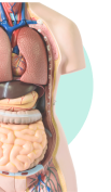
X-Ray scan
X-ray, a form of electromagnetic radiation, is a crucial tool in medical imaging. It enables the visualization of internal structures within the body, aiding in the diagnosis of various medical conditions. This non-invasive technique is commonly used to detect fractures, infections, and abnormalities in organs. By capturing images of the body's internal structures, X-rays provide valuable insights for medical professionals, guiding them in developing treatment plans and monitoring patient health.


Chest X-Ray Scan in Chennai
Looking for an affordable Chest X-Ray scan in Chennai? Bajaj Finserv Health connects you to the best Chest X-Ray scan centers near you, offering accurate results with state-of-the-art technology. Check the Chest X-Ray scan cost in Chennai today and take a step towards better health with competitive pricing and top-notch diagnostic services.
Showing 1 Chest X-Ray Scans in Chennai
Browse by Categories

Full Body Checkups
Overall health diagnosis
Body Organs
Assess major organ function

Lifestyle Habits
Measure habits, empower health
X-Ray, MRI & Scans
Radiology based lab tests
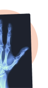
What are X-rays?
X-rays are a type of electromagnetic radiation. These are used in medical imaging to create detailed images of the body's internal structures, including bones, organs, and tissues. X-rays are essential in medical diagnosis as they can diagnose bone deformities and fractures.
How do X-rays work?
X-rays work by emitting a controlled amount of radiation through the body. This radiation passes through the body and creates an image on a detector, which a healthcare professional can then analyze.
Benefits of X-rays imaging
Helps detect and diagnose fractures and bone injuries - Evaluating the condition of the lungs and detecting respiratory issues - Identifying abnormalities in the gastrointestinal system, such as blockages or ulcers - Guiding the placement of medical devices, such as pacemakers or catheters - Assist in the detection and monitoring of certain types of cancer
Safety Considerations and radiation exposure
X-ray procedures are generally considered safe. - The radiation exposure during an X-ray is typically minimal and within acceptable limits. - Pregnant women should inform their healthcare provider before an X-ray examination to ensure appropriate precautions are taken.
Tips for preparing for an X-ray
Follow the instructions given by your healthcare provider or radiology department. - Inform your doctor if you are pregnant or suspect you might be. - Remove any jewelry or metal objects present in the area that is being examined, as they can interfere with the X-ray image.
What happens during an X-ray examination?
You may be asked to change into a hospital gown and remove any clothing or objects that could interfere with the X-ray. - You will be positioned on an X-ray table or stand in front of the X-ray machine. - The technologist will guide you into the appropriate position to obtain the necessary images. - You may need to hold your breath briefly to minimize any motion that could blur the image. - The X-ray machine will emit a brief burst of radiation, which is painless and usually takes only a few seconds. - Multiple images may be taken from different angles or positions. - After the examination, you can usually resume your normal activities unless instructed otherwise by your doctor.

How much does a Chest X-Ray scan cost in Chennai?
If you’re looking for affordable Chest X-Ray scan options in Chennai, Bajaj Finserv Health offers a wide range of diagnostic centers with transparent pricing. The cost of a Chest X-Ray scan can vary depending on the facility and the type of scan. We make it easy to compare prices from different labs so you can find the best option without compromising on quality. Book your Chest X-Ray scan online and get the best rates in Chennai today!
Where can I find a Chest X-Ray scan center near me?
Need to find a Chest X-Ray scan center near you? With Bajaj Finserv Health, you can easily locate trusted diagnostic centers in Chennai for your Chest X-Ray scan. Our platform connects you with certified labs in your area, ensuring that you receive high-quality service and accurate test results, right when you need them. Simply enter your location, and we’ll show you the best options for Chest X-Ray scan near you.
What is the price of a Chest X-Ray scan?
Wondering about the price of a Chest X-Ray scan? The cost of diagnostic tests like Chest X-Ray scan can differ depending on the facility, technology, and location. At Bajaj Finserv Health, we partner with top labs across Chennai to offer competitive pricing, giving you the flexibility to choose the lab that suits your budget without sacrificing quality. Explore options and book your test online for peace of mind.
Factors Affecting Chest X-Ray Scan Price in Chennai
- Lab certification and accreditation status
- Center location and accessibility
- Test methodology and equipment used
- Turnaround time for results
How much is a Chest X-Ray scan in Chennai?
The price of a Chest X-Ray scan in Chennai typically ranges based on the lab’s services and equipment. Bajaj Finserv Health partners with multiple diagnostic centers across the city, making it easy for you to find a reliable Chest X-Ray scan at an affordable cost. Whether you’re looking for standard or advanced tests, we ensure transparent pricing and convenient online booking, so you know exactly what to expect.
How can I book a Chest X-Ray scan in Chennai?
If you need a Chest X-Ray scan in Chennai, Bajaj Finserv Health offers a hassle-free booking process. You can find certified labs near you offering Chest X-Ray scan and other diagnostic services with just a few clicks. Whether it’s for a routine check-up or a specific medical need, our platform helps you find the right lab at the right price in Chennai.
How do I find a Chest X-Ray scan near me?
Searching for Chest X-Ray scan centers near you? Bajaj Finserv Health provides an easy-to-use platform to find and book diagnostic tests in your area. Whether you’re in Chennai or another location, you can locate top-rated labs offering Chest X-Ray scan tests at competitive rates. Simply enter your location, choose a lab, and book your test online for a seamless experience.
Why Choose Our Partner Labs for Chest X-Ray Scan in Chennai?
Our network of trusted diagnostic centers in Chennai ensures reliable Chest X-Ray scans with consistent quality. Enjoy added benefits such as home sample collection, quick access to digital reports, and affordable pricing.
Key Features
- Accredited NABL/ISO certified laboratories
- Access to digital reports for convenience
- Easy online booking and flexible payment options
How much does Chest X-Ray scan cost in Chennai?
The cost of Chest X-Ray scan in Chennai can vary based on the diagnostic center, the type of scan required, and additional factors like the use of contrast material. To get the best deal, visit our platform to check the latest prices and compare offerings from multiple accredited diagnostic centers
How can I find Chest X-Ray scan centers near me in Chennai?
Visit the Bajaj Finserv Health website and explore the Lab section to find accredited Chest X-Ray centers near you. You can compare prices and book appointments conveniently.
What factors affect Chest X-Ray scan prices in Chennai?
Chest X-Ray scan prices in Chennai vary based on scan complexity, equipment quality, use of contrast material, and additional services like consultations and report delivery.
What should I look for in a Chest X-Ray center in Chennai?
Choose a center with NABL accreditation, modern equipment, experienced radiologists, and digital report delivery for reliable results.
How can I book Chest X-Ray scan online?
You can book through our website or app, selecting your preferred center and time slot
What is the cost of Chest X-Ray scan in Chennai?
Prices start from ₹{{min_price}} and vary by center location and facilities.